Magnetic resonance imaging (MRI) is a type of scan that uses strong magnets to generate meaningful signals from the imaged part of our body. These signals are detected by an antenna and processed by a computer to create images (or pictures) of the inside of your body.
Most of the MRI scanners are ‘O’ shaped and appear like a narrow tunnel with a table, on which you lie, slides into the tunnel. Both ends of the scanner are open. Some other MRIs are shaped like a C and other can look like a normal bed (open MRI).
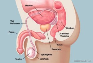
 The prostate gland is a small organ about the size and shape of a walnut, which lies in the pelvis between the bladder and the penis, and in front of the rectum (back passage).
Its function is to help liquefy semen (produced from the male sexual organs to fertilise the female egg).
1. Provide more detailed images of the prostate.
2. To help show if there is any evidence of cancer in the prostate gland if you have a high prostate-specific antigen (PSA) level. PSA substance is produced by the prostatic gland. It is measured by taking a blood sample. It is usually raised when you have prostate cancer, but can also be raised if you have an infection or abscess of the prostate gland
3. To show the extent of prostate cancer within the prostate gland and in the pelvis
4. To help in planning radiotherapy treatment for prostate cancer.
5. To follow up patients who had treatment for cancer prostate to look for recurrence.
You will be asked to complete a questionnaire and safety check. You will then change into a hospital gown and lie on the MRI scanner table. A small needle will be inserted into a vein in your arm or hand to be ably to give you the MRI dye(gadolinium contrast)
The MRI machine produces loud knocking noises, you will either be given earplugs or headphones to listen to music of your choice.
The scan can take 30-40 minutes where you are required to lie as still as possible.
Nothing usually happen from the machine itself.
Some patients develop contrast allergy to MRI contrast which is extremely rare.
Some patients develop rash or allergic reaction.
Small risk of nephrogenic systemic fibrosis – see Contrast Medium: using gadolinium or iodine in patients with kidney problems.
American College of Radiology: www.radiologyinfo.org/en/info.cfm?pg=mr_prostate


Comments
Currently have